By Dr. Ryan Trueman, DC
Pain. It’s a scary word and 95% of the time it is the reason patients step into my office. What many people don’t realize is that pain is a very complex physiological signal that takes into consideration many variables other than structural damage to the body. There are a number of tissues in the body that cause pain when they become tight, dysfunctional, and/or inflamed from both acute or chronic mechanisms. Even your emotions and psychological states can influence your pain and inflammation levels. What’s important to understand is that diagnostic imaging, albeit a wonderful tool in some cases when trying to determine a diagnosis for pain, is not always able to identify the actual cause of it, despite some of the findings that show changes in the body.
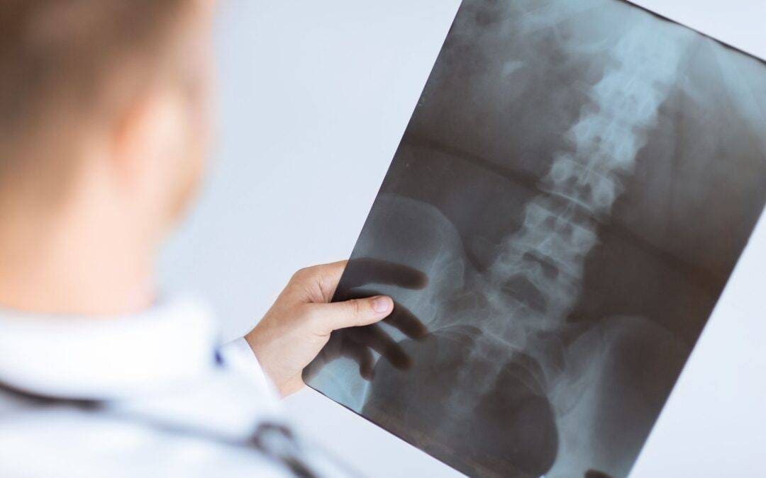
I often meet with patients who have already undergone diagnostic imaging (X-ray, MRI, CT scan, ultrasound, etc.) showing an “abnormal” finding and they are convinced they will not only have pain forever but that their pain is being caused by this finding. The problem here is that often, patients are experiencing pain for a number of different reasons, many of which are not able to be identified by imaging. Just because your X-rays show that you have arthritic joints doesn’t mean that the pain is necessarily caused by arthritis and that you will have to live with pain for the rest of your life. The arthritic findings are from years of wear and tear in the joint and likely represent more of a consequence of the underlying problem than the actual problem itself.
I can’t count how many times I’ve met with a patient for an initial consultation and I’m told something along the lines of “My x-rays showed that I have pain in my knee because of arthritis,” or “I have a disc bulge and degenerative disc disease and it’s causing awful pain in my back.” In reality, your diagnostic imaging is identifying any structural changes in the region being examined and provides us further insight into making a formal diagnosis. In other words, it gives us additional information into one of many possibilities as to what may be causing you pain. When addressing pain due to physical injury or musculoskeletal (MSK) conditions, it’s far more common to be able to make an accurate diagnosis through a thorough history and physical examination than to rely on diagnostic imaging. We use diagnostic imaging if the examination findings aren’t consistent with the history of the problem, if we are concerned about more serious causes of pain, such as infection or tumors, or if the suspected diagnosis requires the imaging to confirm/deny it. The fact is, degeneration in the body is a normal sign of an aging body much like wrinkles on your skin are. My all-time favorite quote on this subject is by Dr. Stuart McGill, a renowned biomechanics researcher and long-time professor at Waterloo researching low back pain. He said, “A degenerative disc disease diagnosis is the equivalent of telling your mother-in-law that she has degenerative face disease!”
Now of course, diagnostic imaging can be incredibly valuable in healthcare and can help direct us from a diagnosis and treatment standpoint if there is structural damage in the tissue. By no means are all findings of degenerative changes on x-rays unimportant. But let’s look at some of the numbers in the research on asymptomatic (no symptoms or pain) patients and their prevalence of having an “abnormal” x-ray/MRI finding. In one study, researchers looked at the estimated percentage of people with x-ray findings for various “degenerative diseases” that function perfectly normally in daily life with no issues. What they found was over 50% of 30-year-olds and almost 90% of 60-year-olds show signs of degeneration on imaging with no symptoms. We know that as you get older almost everyone will have what is considered normal degeneration. This puts in question what is really an abnormal/normal finding and what is clinically relevant to what’s causing the patient’s pain.

Another study on “abnormal” knee findings using MRI in asymptomatic patients in their 50’s, 60’s and 70’s demonstrated similar significant findings. The study found, just to name a few, damage to the cartilage lining of the femur, underlying bone damage, and injury to the meniscus of the knee, with none of these study participants reporting any symptoms . The percentage of knees that showed these MRI findings only increased with every additional decade in age grouping. This once again demonstrates that there appears to be a normal amount of wear that occurs to the knees over a lifetime, yet it doesn’t necessarily correlate with the patient experiencing pain.
Lastly, let’s look at a study on the shoulder where a diagnostic ultrasound demonstrated a rotator cuff tear. Even in the shoulder, there is normal wear and tear on the AC joint of the shoulder (especially those into heavy lifting exercise), the bursa (bursitis), the supraspinatus (1 of 4 rotator cuff muscles and often the most injured), and the subscapularis (another commonly injured rotator cuff muscle/tendon). The study found that 40% of 40-year-olds show a rotator cuff tear but experience no pain. Is this really an abnormal finding and the cause of your pain? In many cases, I have seen older patients that have full-thickness tears (torn completely) and are still not exhibiting any pain or dysfunction.

So if what I am seeing on my x-ray or MRI isn’t causing my pain, what is?
To answer this we have to answer the question what is pain. Pain is a signal or an alarm, that’s it. It comes in many different forms and much like your car alarm can go off when there is no REAL threat, our bodies can sound the alarm when there is no damage or threat as well.
Most people think of pain as a signal that we are doing damage to our bodies, but this is not the only trigger for pain. For example, pain can come from weak and deconditioned tissues not able to cope with our day-to-day requirements, excessive tension in muscles and fascia (connective tissue) where repetitive movement creates stress and tissue inflammation, and of course from acute injuries such as contusions (bruises), sprain/strains from sports-injuries, and tissue loading problems such as lifting injuries in the low back. The commonality between all of these examples? Most, if not all of these types of injuries will likely not be specifically identified on imagining other than the consequential inflammation associated with the injury.
As previously mentioned, emotional pain associated with depression/stress/anxiety can cause physical pain and has been shown to cause both chronic (long-term) and acute inflammation (our body’s reaction to injury). Even FEAR of pain can even cause physical pain! The very act of thinking “I can’t deadlift anymore because I have degenerative disc disease and it is always going to hurt” can actually bring on pain before you even perform the lift!
To summarize by example, the majority of x-ray findings you will often see from longstanding wear and tear in the spine doesn’t mean you have to live with low back pain for life! In fact, more often than not the pain-generating structures will have nothing to do with what’s seen in your diagnostic imaging. It’s possible your degenerative disc disease isn’t causing your pain, but rather the muscular imbalance of your core stabilizers or the longstanding lack of hip mobility from fear of using your spine! The overall message here is that I encourage you to not let imaging findings be the only piece of information that tells you what your underlying problem is. Work with a healthcare professional who can perform a thorough history and physical examination, and if necessary, use diagnostic imaging in conjunction with the clinical findings to properly advise you on the problem and the appropriate solution. As per my favorite quote by Dr. McGill, make sure to focus on the whole picture and not a just snapshot of the wrinkles on your face.